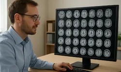
Radiology has long been a bottleneck in brain care. MRI scanners can generate hundreds of images per patient, and interpreting them demands time, specialized training, and careful cross-checking against clinical history. When the stakes include stroke, tumors, neurodegeneration, or inflammatory disease, speed matters-but so does precision.
Researchers at the University of Michigan have described an AI model designed to do something ambitious: diagnose more than 50 brain disorders from MRI scans in seconds. The work, published in Nature Biomedical Engineering, reports performance reaching up to 97.5% accuracy in testing, positioning the system as a potential assistant for clinicians facing growing imaging volumes and complex differential diagnoses.
The headline number is attention-grabbing, but the deeper story is about how AI is being shaped to fit the realities of neuroimaging: messy data, overlapping disease signatures, and the need for tools that can generalize beyond a single hospital's scanner settings.
Why brain MRI is hard to "automate"
MRI is one of the most information-rich tools in medicine. It can highlight anatomy, blood flow, tissue damage, and subtle changes in white matter. That richness is also the problem. Different MRI sequences emphasize different tissue properties, and protocols vary across institutions, scanner vendors, and even individual radiology departments.
Many brain disorders also share imaging features. Swelling, lesions, atrophy, and signal changes can appear in multiple conditions, and the same disease can look different depending on stage or patient-specific factors. Radiologists and neurologists often combine imaging with symptoms, lab tests, and patient history to narrow down possibilities.
Traditional computer-aided detection tools have typically focused on narrow tasks-spotting a tumor, measuring brain volume, or flagging a bleed. A model that attempts to classify dozens of disorders at once is trying to mirror the breadth of real-world diagnostic work rather than a single, isolated pattern-recognition problem.
What the University of Michigan model is aiming to do
According to the report, the University of Michigan system can diagnose more than 50 brain disorders from MRI scans in seconds. That scope suggests a multi-class approach: instead of asking the model to answer one question ("Is there a tumor?"), it is asked to choose among many possible conditions.
The study's reported accuracy-up to 97.5%-is a measure of performance under the researchers' evaluation setup. In medical AI, that context matters. Accuracy can vary by disorder, by imaging protocol, and by how balanced the dataset is across conditions. A system can score highly overall while still struggling with rarer diseases or borderline cases.
Still, the combination of speed and breadth is the point. If a model can rapidly triage scans and suggest likely diagnoses, it could help clinicians prioritize urgent cases, reduce time to treatment, and standardize interpretation across settings where neuroradiology expertise is scarce.
How AI "sees" an MRI: a practical technical view
Most modern medical imaging AI relies on deep learning, often using convolutional neural networks or related architectures adapted for 3D data. Brain MRI is not a single picture; it is a volume. That means the model must learn spatial relationships across slices and across sequences.
In practice, an MRI-based diagnostic model typically has to handle several steps:
- Preprocessing and normalization: Aligning scans, standardizing intensity ranges, and reducing scanner-specific quirks so the model doesn't learn the wrong cues.
- Feature learning: Identifying patterns correlated with disease-lesion distributions, tissue changes, structural asymmetries, or diffuse atrophy.
- Classification: Producing probabilities across many disorders, sometimes with an "uncertain" or "other" output if the scan doesn't match known categories.
- Quality control: Detecting motion artifacts or incomplete sequences that could mislead the model.
A key challenge is generalization. A model trained on one set of scanners can fail when it encounters different acquisition parameters. Research teams often address this by training on diverse data, using augmentation techniques, or applying domain adaptation methods that reduce sensitivity to site-specific differences.
The University of Michigan work is presented as a step toward that broader, more clinically useful kind of model-one that can handle a wide diagnostic menu rather than a single condition.
Speed is not just convenience-it changes workflow
"In seconds" is more than a performance brag. It points to a different way imaging could be used in care pathways. Today, MRI interpretation often sits in a queue. Even when radiology departments are efficient, the process includes scan acquisition, transfer to PACS systems, reading, dictation, and report distribution.
An AI system that can generate a preliminary assessment quickly could be used for triage. For example, it might flag scans that look consistent with time-sensitive conditions, pushing them to the top of a radiologist's worklist. It could also provide a second read, highlighting cases where the model's output diverges from the initial human interpretation.
That said, speed can create its own risks. If a fast model is treated as authoritative, it may encourage overreliance. The more realistic near-term role is assistive: a tool that helps clinicians move faster while keeping humans responsible for final decisions.
What "97.5% accuracy" can-and can't-tell you
Medical AI performance metrics are notoriously easy to misunderstand. Accuracy is a single number that compresses many realities: false positives, false negatives, class imbalance, and the clinical cost of different errors.
For brain disorders, the consequences of mistakes vary widely. Missing a rapidly progressing condition is not the same as misclassifying two disorders with similar management. A model's usefulness depends on how it performs per condition, how it behaves on borderline cases, and whether it can communicate uncertainty.
The study's reported peak accuracy is a strong signal that the approach is working under test conditions. The next questions for any such system are about robustness: performance across hospitals, across patient populations, and across real-world imaging variability.
Clinical adoption: integration is the real hurdle
Even high-performing imaging AI can stall when it meets hospital IT. To be used routinely, a diagnostic model must integrate with existing radiology infrastructure-PACS, reporting systems, and electronic health records-without adding friction.
There are also operational questions. Who is responsible for monitoring model drift over time? How are software updates validated? What happens when the scanner protocol changes? Hospitals need governance processes that treat AI like any other clinical system: monitored, audited, and maintained.
Regulatory clearance and clinical validation are separate steps from publishing a strong research result. A model can be impressive in a paper and still require extensive prospective testing before it is trusted in routine care.
Implications for radiologists and neurologists
AI in radiology is often framed as replacement versus augmentation. In practice, the most plausible impact is redistribution of effort. If AI can handle broad screening and pattern recognition, clinicians can spend more time on complex cases, correlating imaging with symptoms, and communicating with patients and care teams.
For neurology, a tool that suggests a ranked list of likely disorders could shorten the path from symptoms to diagnosis, especially in settings where subspecialty expertise is limited. It could also encourage more consistent interpretation across institutions, reducing variability that comes from differences in training or workload.
At the same time, multi-disorder models raise new expectations. If a system claims to cover dozens of conditions, clinicians will want to know where it is strong, where it is weak, and how it behaves when confronted with rare diseases or mixed pathologies.
The broader industry trend: from single-task AI to "diagnostic platforms"
Early medical imaging AI products often targeted one narrow use case because it simplified training, validation, and regulatory pathways. The University of Michigan model reflects a shift toward broader systems that can support differential diagnosis across many conditions.
That shift has implications for how imaging AI is built and sold. A multi-disorder model could reduce the need for hospitals to manage a patchwork of single-purpose tools. It also raises the bar for transparency and evaluation, because a single system may touch many clinical decisions.
If research like this continues to translate into deployable software, the competitive landscape could move toward platforms that combine imaging interpretation with workflow tools-triage, reporting assistance, and longitudinal tracking-rather than isolated algorithms.
What to watch next
The University of Michigan study adds momentum to the idea that AI can do more than detect one abnormality at a time. The most important next steps are likely to revolve around real-world performance: how well the model handles diverse scanners, varied patient populations, and the full range of clinical complexity.
Clinicians and hospital leaders will also look for clarity on how such a system communicates uncertainty, how it can be audited, and how it fits into existing diagnostic pathways without creating new safety risks.
For patients, the promise is straightforward: faster answers from scans that already exist in the care process. The challenge is making sure those answers remain reliable when the model leaves the controlled environment of a study and enters the noisy reality of everyday medicine.


